Hip pain is very common phenomena in our society, Since the hips carry the gravity center in our body, they form the foundation of the all body. Every muscle above and under the hips will have to respond automatically to every distortion or change in the location of the hips. To enable the body to function in the most efficient way the hips have to be leveled horizontally.
We will look at the most common bodily distortion and its effect on the body in the reclining position, while standing and while walking. In the most common bodily distortion, the right hip is rotated forward approximately ¼ inch to 1 inch more that the left hip. This occurs because there is frequently a tendency to use the right leg more than the left leg, for instance, as in the operation of an automobile with automatic transmission. In this case, use of the right leg activates the muscles of the right hip, causing these muscles to shorten and consequently to pull the hip forward. The effects of this mechanical distortion may be clearly seen by examining the subject in various positions.
The supine (reclining) position: When the subject lies on his back, the right leg will extend further forward than the left leg, making the right leg appear longer that the left leg. This is not because of an anatomical discrepancy in leg length, but because the right femur (thigh bone) extends from the right hip which, in our subject, is rotated forward.
In the standing position: When the subject is standing he will automatically shift his weight onto the functionally shorter left leg. This tilts the pelvis to the left, pulling the spine and head also to the left. This change in head position will involve the righting reflex, which will be called into action to reestablish the eyes on the horizontal plane.
This neuromuscular reflex will be carried out as follows: The nervous system will send impulses to muscles on the right side of the trunk to contract, thereby pulling the spine and head to the right, as a reaction to the left tilt of the hips. Next, the nervous system will recruit muscles between the left shoulder and neck to pull the head back to center to level the eyes. While the subject is standing, his right hip will be higher than the left hip, and his left shoulder will be higher than the right shoulder. Most people fit this pattern. The simple act of standing results in excessive muscular activity that distorts skeletal alignment, all in reaction to the initial simple forward rotation of the right hip.
Every muscle above and under the hips will have to respond automatically to every distortion or change in the location of the hips.
The person may not suffer pain at the moment, but eventually this constellation of distortions in skeletal alignment will result in some degree of chronic debility and pain. The muscles which are involved in hip pain are:
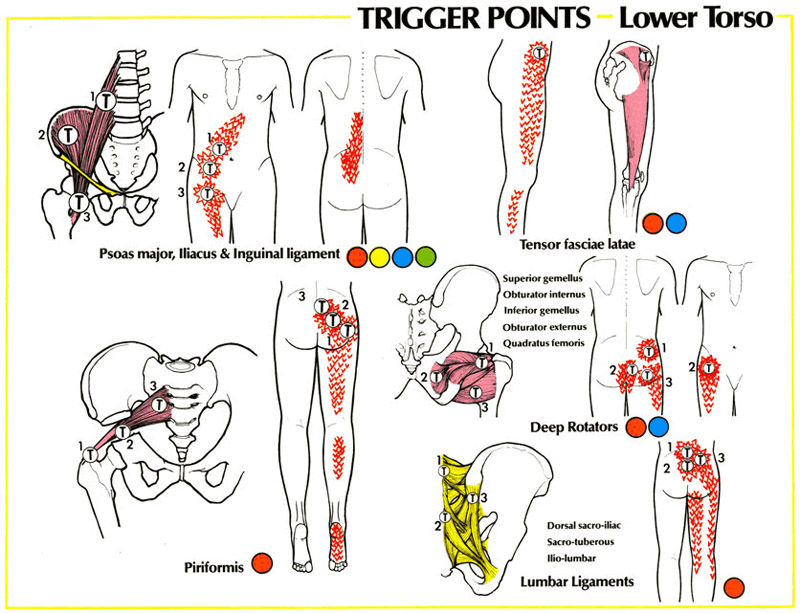
- Psoas – Extremely important muscle, attached to the spine from the front and side, and connected to the inside of the hip and leg. This muscle is the main culprit of lower back and hip pain in our society, since we have become very sedentary. This muscle is the most used and abused in the body, because we use it all the time, while walking, standing and sitting. When this muscle gets tired and spastic, it will pull on the femur (thigh bone) onto the accetabulum (the hip socket) and increase the pressure on the hip joint. If the pressure lasts for a long time it will cause deterioration of the cartilage of the hip joint and hip replacement will be unavoidable when this muscle is spastic it will refer pain to the lower back, the front of the hip and leg.
- Piriformis – This deep muscle is located in the center of the buttocks. It originates in the front side of the sacrum (tail bone) and connects to the hip joint. When the piriformis is in a spasm it will compress the thigh bone into the hip socket increasing the pressure on the joint. Also, when this muscle is spastic it will compress different nerves roots which exit the Sacrum (tail bone), including the “famous” Sciatic nerve. The piriformis muscle syndrome is frequently characterized by such bizarre symptoms that may seen unrelated. One characteristic complaint is a persistent severe, radiating low back pain extending from the sacrum (tail bone) to the hip joint, over the gluteal region and the posterior of the upper leg, and behind the knee. In the most severe cases the patient will be unable to lie or stand comfortably, and changes in position will not relieve the pain. Intense pain will occur when the patient sits or squats since this type of movement requires external rotation of the upper leg and bending the knee.
- When the Sciatic nerve is involved the patient will experience also tingling and numbness in the calf and foot.
- Tensore Fasciae Latae – This small muscle on the side of the hip connects the hip bone to the femur (thigh bone) from the side. Since 90% of the time during walking the body weight is carried by one hip, the hip muscles are forced to work harder which make them susceptible to get in a spasm. When this muscle is spastic, pain would be referred to the side of the thigh and calf. It would be very painful, sometimes impossible to put any weight on the hip.
- Gluteus maximus, medius and minimus – These superficial hip muscles cover the whole hip from the waist to the hip joint itself. When they are spastic , pain would be felt in the hip and referred to the back and side of the thigh and calf. It would be very painful, sometimes impossible to put any weight on the hip.
- Deep Rotators – These five deep muscles connect the hip joint to the bottom of the pelvis from the back. When they are in a spasm the pain would be felt in the hip and sacrum (tail bone). Sitting would be very uncomfortable.
- Lumbar Ligaments – These three ligaments connect the hip joint to the lumbar spine (low back) and the sacrum (tail bone). When they are spastic, pain would be felt in the low beck hip and would be referred to the back of the thigh.
Hip Pain Referral Patterns
Hip Pain Referral Patterns 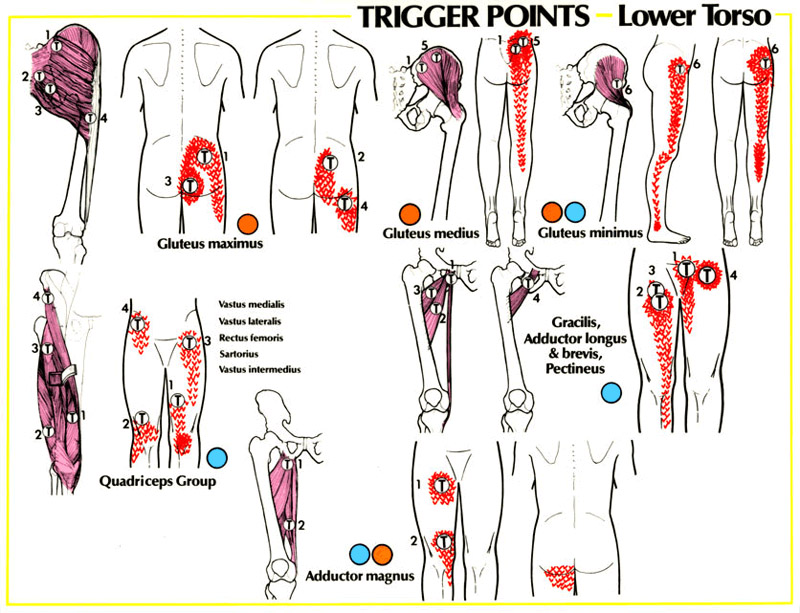
Hip Pain Referral Patterns