Knee pain could be affected by the unevenness of the hips. Since the hips carry the gravity center of the body, this center will be thrown off causing the legs length not to be equal, and as a result of it more weight will be loaded on one knee than the other.
If this condition lasts, it may cause a spastic reaction in different muscles, which support the knee.
These muscles could be in the front, side or back of the leg. Only a proper diagnosis can determine the cause of the pain.
These pain patterns frequently present in older patients who already have some degenerative arthritis and may have been told they will ‘have to live with it.’
Pain in the anterior medial knee:
- Rectus femoris – This muscle connects the hipbone to the kneecap and is very common myofacial syndrome.
- Vastus medialis – This muscle connects the hipbone to the inside of the kneecap. Also is very common myofascial syndrome.
- Adductor longus and brevis – These muscles connect the hip bone to the inside of the thigh.
Patients usually experience anterior knee pain getting up from a seated position. Often it is necessary to push up with their arms to be able to stand. Getting upstairs is also difficult and painful. Any activity that stresses or stretches the three muscles listed will cause discomfort. When pathology in these muscles is the cause of the knee pain, treatment of the Tender Points will provide immediate relief. The patient should be able to perform previously painful maneuvers without pain before the visit is over. Mayofascial pain is very amenable to counterstrain manipulation, and results in a very grateful and astonished patient. These pain patterns are frequently present in older patients who already have some degenerative arthritis and may have been told they will “have to live with it.” This is not true. You can find relief.
Pain in the lateral knee:
- Vastus lateralis – This muscle connects the hipbone to the outside of the thigh and kneecap and is the most commonly found myofascial pain pattern in the lateral thigh.
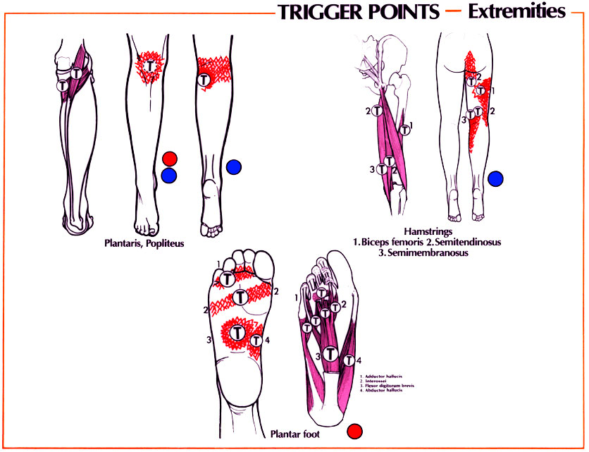
Pain in the posterior knee:
- Gastrocnemius – This muscle connects the back of the thigh to the heel of the foot and is the most common myofascial pain pattern in this area. Patients present with pain in the posterior knee, especially when the knee is extended (straight) and the ankle is flexed (bent).
- Biceps femoris – This muscle connects the hipbone from the back to the lateral upper side of the calf and is the kneenext most common pain pattern. Often seen in patients with athletic injuries that present with posterior knee pain.
- Soleus – This muscle connects the upper back part of the calf to the heel of the foot and is also common, but usually has more posterior calf pain than pain in the knee.
- Semimembranosus and semitendinosis – These muscles connect the hipbone from the back to the medial upper of the calf.
- Popliteus – This muscle connects the lower lateral side of the thigh to the upper lateral side of the calf.
Knee Pain Referral Patterns
Knee Pain Referral Patterns 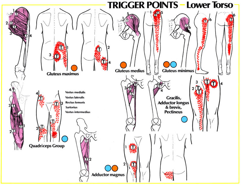
Knee Pain Referral Patterns 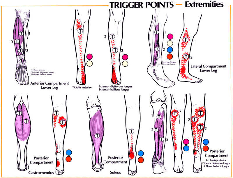
Knee Pain Referral Patterns